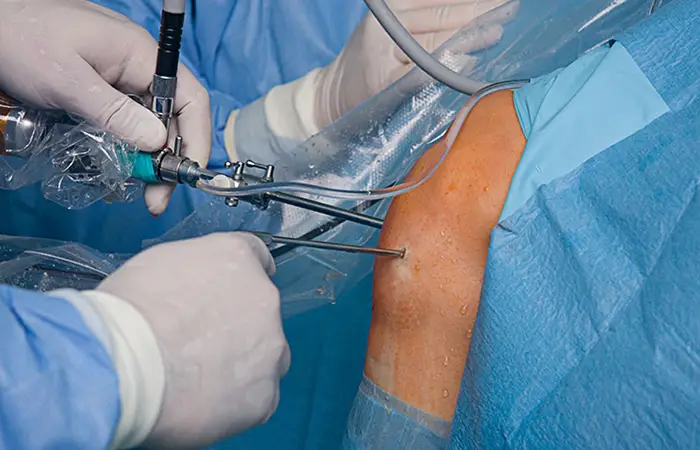
Arthroscopy (ahr-THROS-kuh-pee) is a procedure for diagnosing and treating joint problems. A surgeon inserts a narrow tube attached to a fiber-optic video camera through a small incision — about the size of a buttonhole. The view inside your joint is transmitted to a high-definition video monitor.
Arthroscopy allows the surgeon to see inside your joint without making a large incision. Surgeons can even repair some types of joint damage during arthroscopy, with pencil-thin surgical instruments inserted through additional small incisions.
Doctors use arthroscopy to help diagnose and treat a variety of joint conditions, most commonly those affecting the:
Diagnostic procedures
Doctors often turn to arthroscopy if X-rays and other imaging studies have left some diagnostic questions unanswered.
Conditions treated with arthroscopy include:
Arthroscopy is a very safe procedure and complications are uncommon. Problems may include:
Exact preparations depend on which of your joints the surgeon is examining or repairing. In general, you should:
The type of anesthesia used varies by procedure:
One small incision is made for the viewing device. Additional small incisions allow insertion of surgical tools.
Your aftercare may include:
Call your surgeon if you develop:
In general, you should be able to resume desk work and light activity in a few days. Driving may be possible in one to three weeks. Full recovery depends on your condition.
Your surgeon will review findings and monitor progress during follow-up visits.